Images of the Nucleosome
From the X-ray crystal structure of Richmond and colleagues: Luger, K., Mäder,
A. W., Richmond, R. K., Sargent, D. F. & Richmond, T. J. (1997). Crystal
structure of the nucleosome core particle at 2.8 Å resolution. Nature
389, 251-260.
Here is the PDB file, 1AOI.PDB. And here is a Rasmol
command file, as usual, suitable for cutting and pasting into the Mac version.
This version of the command file has pause statements for running as
a script in Windows.
Here is a Jmol tutorial that uses the more recent 1KX5 structure.
You should get results approximately like these images:
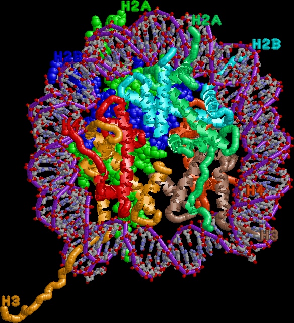
- An overview of the whole nucleosome (except for much of the tails, which
were disordered). Histones H3 and H4 are in earth tones, H2A and H2B in
sea tones. Note that the (H3/H4)2 tetramer binds both gyres near the dyad
axis, whereas each H2A/H2B dimer binds mainly one gyre.
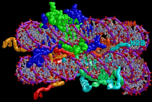
- A view from the side, showing that the H2A/H2B dimers don't have much contact with each other,
and again contact mainly one gyre.
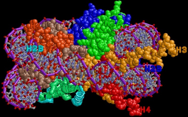
- A view from the side looking at the nucleosomal dyad, emphasizing how the H3/H4 tetramer holds everything together.
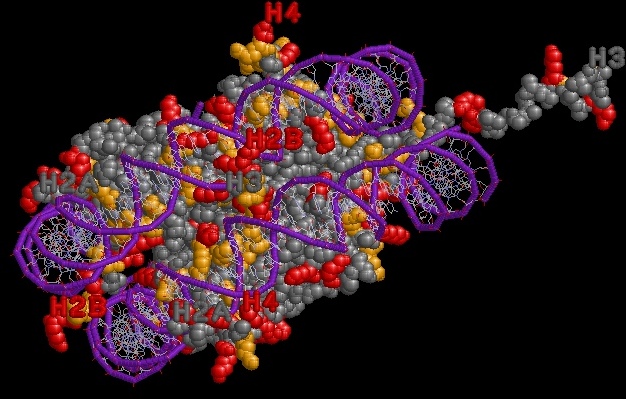
- Arginine in orange, lysine in red. Note the many arginines making minor
groove contacts to DNA, and the concentration of positively charged residues
where the DNA binds.
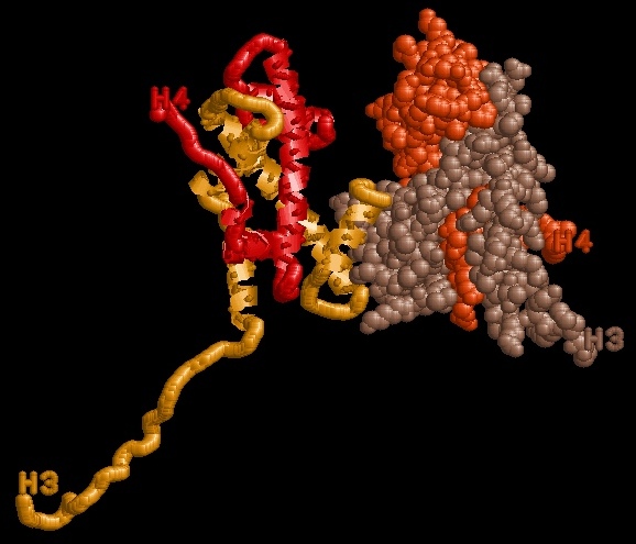
- A closer look at H3/H4. On the left, the histone fold dimerization motif holds the gold and red
H3 and H4 together. The tetramer interface is a four-helix bundle formed between the two H3's.
Back to J. Kahn's Teaching Resources.
Or my home page.
Last modified 4/6/02.