This page is a skeletal tutorial on the structures of A and B form
nucleic acids and on the use of the Jmol plugin to display
three-dimensional structures (here, "PDB files", where PDB = protein data bank
format).
|
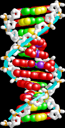
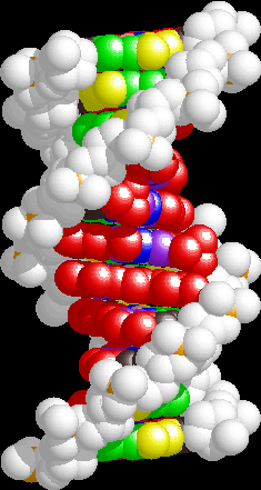
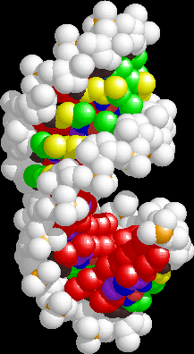
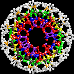
Here are gif images generated from the idealized A and B form PDB files used
above, made using Rasmol, which is an earlier stand-alone incarnation of Jmol.
The images in the left two columns emphasize the major and minor grooves
of the DNA and RNA. The left column shows "wireframe" images. The center column
shows spacefilling images. The
color coding is as follows:
|
Major groove -- red
Minor groove -- yellow
|
Watson-Crick H-bonding face -- blue
major groove and WC face -- purple
major groove and WC face -- green
|
backbone -- white, with orange P atoms
other atoms -- brown. Sugar protons not shown.
imino protons -- pink
|
|
B-DNA. The helix backbone is in cyan.
|
B-DNA. Note the large, accessible major groove and narrow minor
groove.
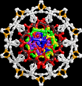
|
The top view shows the base pairs in the center of the helix.
|
A-form RNA. Note the inclined base pairs.
|
A-RNA. Note the deep, inaccessible major groove and shallow minor
groove.
|
A-RNA. Note the base pairs are offset toward the outside of the helix.
|