Here are images made in Rasmol as described in the referring
page.
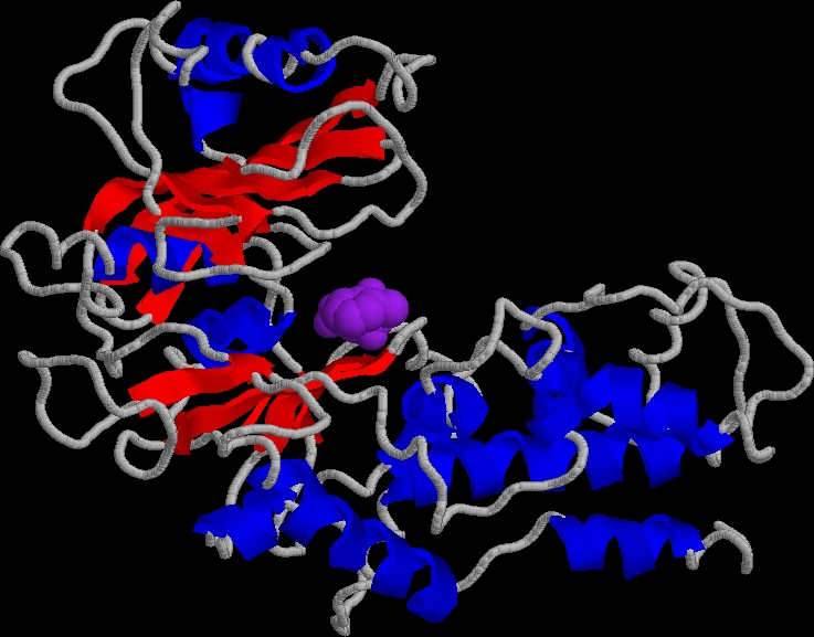
The open from (2yhx.pdb). The inhibitor OTG is shown in purple:
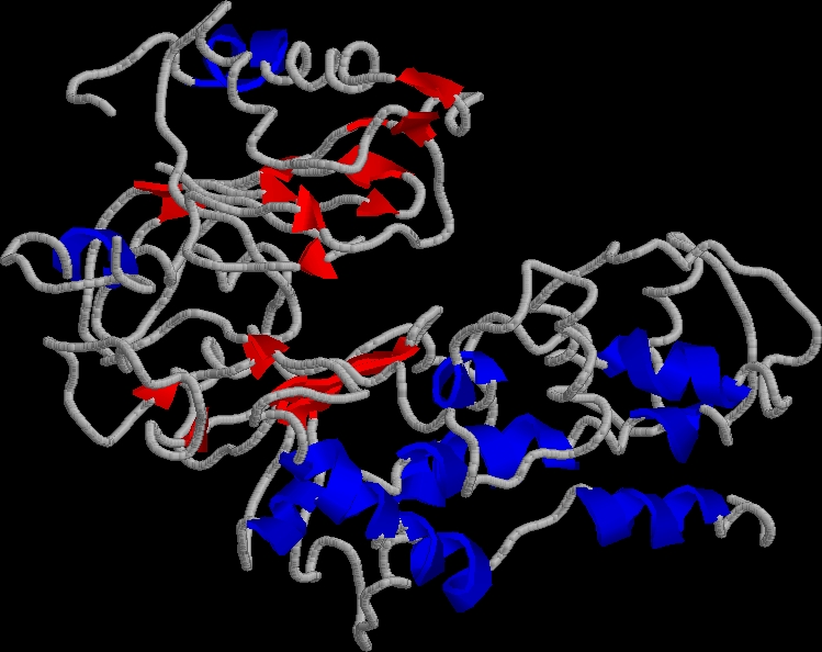
The closed form (1hkg.pdb). Note that the glucose substrate was not visible in the X-ray structure. The textbook pictures model it in from the position of the OTG inihibitor in the open form.